

Digital X-Rays
X-rays are an essential diagnostic tool for dentistry. The ability to visualize the density of teeth and the bone that supports the teeth gives so much more information than we would be able to gather without x-rays.
How does it work?
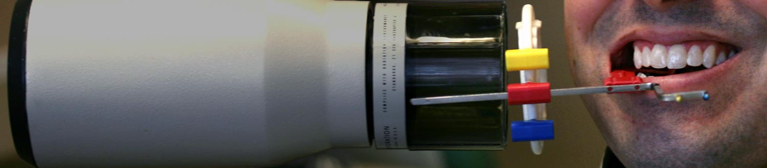
Put simply, x-rays work by focusing a beam of radiation through a targeted area and onto a sensor that detects the amount of radiation that was allowed through the area being targeted. Dense objects like teeth will shield the sensor from the radiation and will appear white (or radio-opaque) on the image produced. Less dense objects like air or cavities show up as black (or radio-lucent).
Types of X-Rays
Bitewings
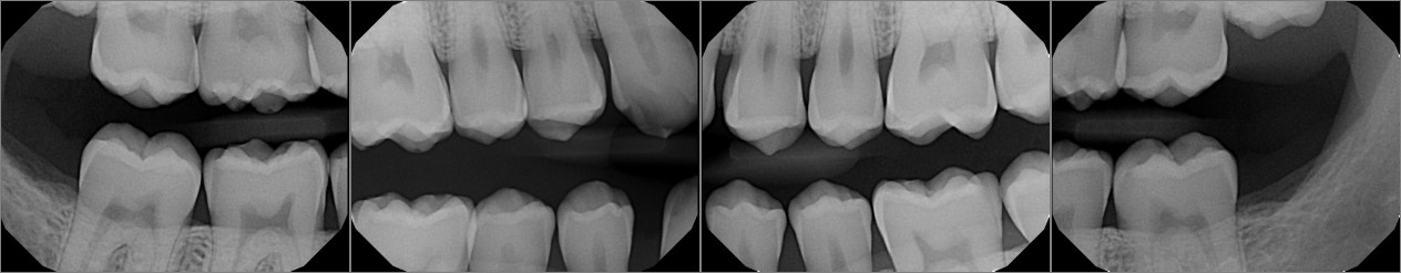
This is a typical set of x-rays that you may receive at your routine dental visits. They are the most useful type of x-ray for diagnosing caries (AKA tooth decay or “cavities”). We can also see calculus (“tartar”), bone height, previous restorations, and other abnormalities. With bitewing x-rays we cannot see the roots of the teeth or the anatomy surrounding the roots. These are also only needed for viewing the back teeth, since the front teeth are thin enough that we can usually see cavities without x-rays. We will typically take 4 bitewings for adults, and 2 for children. Taking these periodically helps us to monitor for early signs of cavities and treat them before they become a problem.
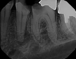
Periapical (PA)
Periapical means “around the apex of the tooth” or around the root. These x-rays are focused on a specific tooth or area to show the entire tooth including the root. This allows us to diagnose any pathology or infection that may be occurring at or near the tooth. These are usually taken if there are symptoms or a problem noticed, or simply to monitor previous root canals or dental implants periodically. We also may use these x-rays to confirm there is no underlying problem with the tooth before we restore it with a large filling or a crown.
Occlusal
This view is helpful with children in order to screen them for any potential problems with their developing adult teeth. It also shows a periapical view of the front teeth.

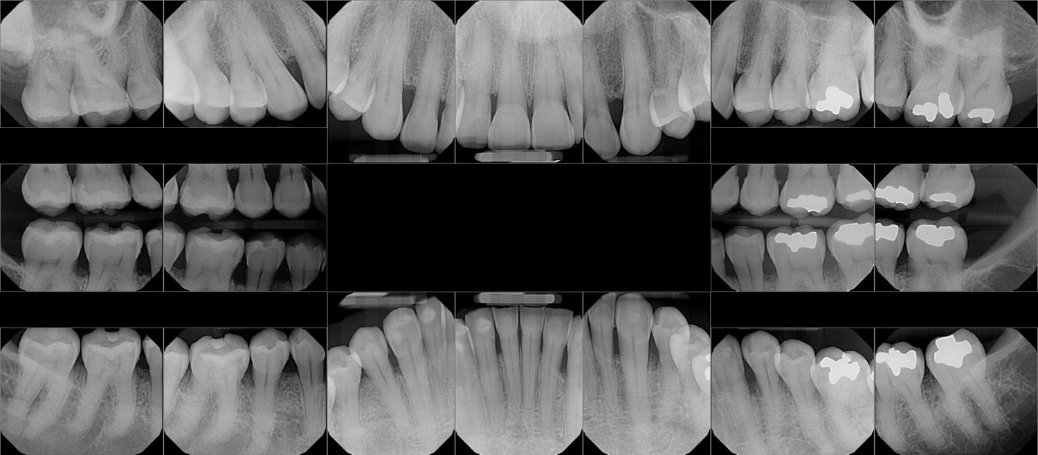
Full Mouth Series (FMX)
An FMX is a series of 18-20 x-rays, which includes all 4 bitewings and PA’s of every tooth. It allows a comprehensive look at every tooth, and is especially useful if there are multiple teeth with problems to diagnose. An FMX, if needed, will usually be used at initial visits to a new dentist, and after 5 years it may be necessary to update.
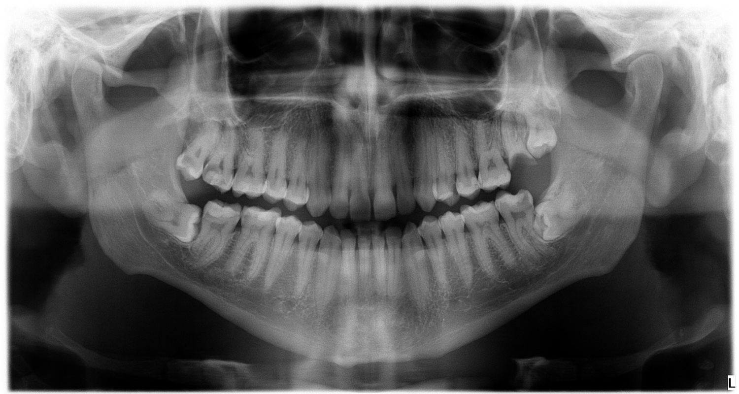
Panoramic
A panoramic x-ray is taken by a machine that rotates around your head to produce an image that includes the teeth, jaws, and sinuses. It is a 2-dimensional image that shows more information than an FMX, but it is not as detailed and is not diagnostic for certain problems like caries. It can be useful for detecting other pathology or in planning for orthodontics or implants.
Cone Beam CT (CBCT)
This is a 3-dimensional scan that is very useful for implant planning and endodontics (root canals). It is similar to a CT scan, however it is different in the method of exposure and is able to use significantly less radiation than a traditional CT scan. The scanner exposes slices from three different angles and is able to reproduce a 3D model of the targeted area. We can also scroll through slices to view bone contours, or root canal morphology that would otherwise be impossible to see with other x-rays. There are many more capabilities, much more than traditional x-rays, however because of higher radiation exposure and higher cost we limit using this technology to those cases that require it.
Note: We do not currently have a CBCT or Panoramic unit in our office. However, we refer to specialists who do have and use them.
Safety
As with all things in healthcare we need to weigh the benefit vs. the risk. There is absolutely a risk when it comes to exposing any part of your body to ionizing radiation, and that is why so many precautions are taken with dental x-rays to limit the amount and frequency of radiation exposure. The beam of radiation is highly calibrated and measured to give the smallest dose of radiation necessary to provide the most benefit. We also place a lead-lined apron over your body to cover your vital organs from any radiation scatter.
Without x-rays it would be very difficult (if not impossible) to diagnose decay between your teeth before it becomes a much larger problem. For this reason, dentists use the ALARA principle (As Little As Reasonably Achievable) when it comes to ordering x-rays. Some people have a low risk for problems and can wait 2 or even 3 years between x-rays. However, many people are at a medium or high risk for problems and should be monitored with at least annual x-rays. People at risk may include those with multiple existing restorations, small areas of decay being monitored, poor oral hygiene, missing teeth, certain medical conditions, etc. It is important to ask your dentist if you really need x-rays, and it is your decision to have them or not, but in many cases the benefits of x-rays outweigh the minimal risks involved.
For more information: Simple Steps to Better Dental Health - X-Ray Frequency Guidelines
Do I need x-rays? I don't feel anything wrong?
See the answer above, but also keep in mind that most cavities are not painful or even noticed until they become very big. At that point, they may need a root canal instead of a simpler and cheaper filling.
The cost is actually significant – a filling may cost around $100-300, and a root canal alone may be around $700-1200. Additionally, most root canals need a build-up and crown so the total cost to restore the tooth may be around $2000-2500, and many people might opt to pull the tooth instead.
If a dentist recommends x-rays, it is in your best interest to accept, however it is certainly your right to refuse. If your hesitation is the cost, please at least consider the cost analysis presented in the previous paragraph, but if you have concerns about the radiation, please continue reading.
Dosage of Radiation
Dosage is important in determining the relative risk or benefit of any medical intervention. It can determine the difference between safe and poisonous with almost anything we ingest (even water). Radiation dosage is similar. Dental x-ray doses have always been fairly small, and now with digital sensors the dose is actually a lot smaller (when we switched to digital sensors we decreased our exposure time to about 40% what it was with conventional films). There are different measurement units for radiation, but the most commonly used in healthcare is the Sievert (Sv), which measures the absorbed dose. Typically we use much smaller units like milli-sievert (1,000 mSv = 1 Sv) or micro-sievert (1,000,000 μSv = 1 Sv) since the dosage is normally very small.
It is also worth noting that radiation is not only caused by man-made objects like medical/dental x-ray imaging. It is also found naturally in our environment. Sources for natural (background) radiation include cosmic radiation from space, terrestrial radiation from the ground, radon in the air we inhale, and small amounts of radiation in food and water sources. These sources all vary depending on where we live, but the average natural dose is something we can use for comparison with the doses of diagnostic tests.
The following chart compares the radiation dose from dental diagnostic x-rays to other sources of radiation in order to give perspective on the relative amounts of radiation exposure. (Disclaimer: Various references were used to gather this information and these are only approximate estimates due to many variables that can factor into these measurements.)
| Dental Radiation Sources | μSv (micro-sieverts) |
|---|---|
| 1 Dental x-ray | 1-2 |
| 4 Bitewings | 5 |
| 16-20 Full-mouth series (FMX) | 35 |
| Panoramic x-ray | 17-26 |
| Cone Beam CT | 12-1000* |
| Other Radiation Sources | μSv (micro-sieverts) |
|---|---|
| Medical Chest x-ray | 100 |
| Medical Chest CT (Standard) | 7000 |
| Eating one banana | 0.1 |
| Daily environmental exposure | 10 |
| Annual environmental exposure | 3100-4000 |
| Annual dose from natural Potassium in the body | 390 |
| Flight from LA to New York (7hrs) | 20-40 |
| Annual dose for a flight attendant | 1500 |
| Exposure Limits | μSv (micro-sieverts) |
|---|---|
| Dose limit for 1 year of occupational exposure | 50,000 |
| Fatal dose (from a single event) | 8,000,000 |
For more information on radiation measurements:
Xkcd - Radiation Dose Chart
ADA - X-Rays
Wikipedia - X-ray Units of Measure and Exposure
RadiologyInfo.org - Radiation Dose in X-ray and CT Exams
Effective Doses in Radiology and Diagnostic Nuclear Medicine (2008)
XrayRisk.com
Health Physics Society
Pregnancy
If you are, or think you might be pregnant it is important to inform us when you come in for your appointment. X-rays are very safe due to the low dosage and shielding provided by the lead apron, and they have never been shown to cause any harm to a developing fetus. However, we still consider the risk vs benefit and usually will postpone routine x-rays unless there is a valid reason. Therefore, if you are symptom free and are at low risk for tooth decay we will most likely wait until after you have the baby to take x-rays. On the other hand, if you have problems such as pain in your teeth and gums or obvious deep cavities, it would be beneficial for us to take the needed x-rays in order to treat your disease. It is important to maintain your health and well-being so you can focus on the developing baby inside you. It is generally believed that the second trimester is the safest time for x-rays and anesthetics, but the risk to the baby is so low that the best time to treat a problem is as soon as possible to avoid any stress or discomfort.
For further information on x-rays during pregnancy, here are some statements from other official organizations:
ACOG (American Congress of OB/Gyn’s): Dental X-rays, Teeth Cleanings = Safe During Pregnancy
Mayo Clinic: Is it safe to have an X-ray during pregnancy?
Mouth Healthy by ADA: X-rays
If you have any questions or comments on this topic, please contact us for more information.